
Wednesday 7th February 2018 – Guy’s Hospital – Haemophilia Clinic
I needed to be up in London by half past nine for a Haemophilia Clinic, even though I’m not a haemophiliac. The first I knew about this appointment was via a text message, just before Christmas, followed up, a few days later, by a letter. On arrival I had my blood pressure and pulse rate measured then settled down with a book, expecting a long wait. A short time later my name was being called.
I was greeted by a doctor I hadn’t met before. After the initial pleasantries she asked “Do you know why you are here?” Tempting as it was to reply “Do any of us? Are we the creation of some omnipotent deity or the product of thousands of years of evolution?” thus proving yet again that I can do pretentious, I opted for “No”. Although tempered this with “… “it’s probably to do with a bleeding management plan”. Correct. It was a follow-on to the meeting with the surgeon to discuss having my gallbladder removed and his concerns about carrying out the surgery.
I don’t want to sound dextraphobic but when I saw that the doctor was left handed I knew it would be a good consultation. We went through my medical history. She was under the impression that I had undergone a major Crohn’s flare in June 2012 so I was able to correct her and explain the incident when my esophageal varices burst. She asked how the problem manifested itself. I replied “sitting surrounded by a pool of blood”.
Previously I had been told that Crohn’s patients undergoing a flare are more susceptible to blood clots but I didn’t understand why. She explained that during a flare the blood becomes extra “sticky” to combat the inflammation. The portal vein carries blood from the gastrointestinal tract and is a common place for a clot to form. The body compensates for the blockage by growing new veins (varices) around the clot but a back-pressure can build up which in turn causes the spleen to enlarge and, in my case, varices to grow around the gallbladder. They would be an added complication should I need a cholecystectomy.
The enlarged spleen stores more platelets rather than release them into the bloodstream. Combine this with the damage to my bone marrow, probably due to Azathioprine, and it explained why blood tests showed my platelets were well below the optimum range. Many patients did not notice they have a problem until the count falls to single figures. Mine were between 60 and 80 ad was perfectly respectable for surgery or dental work. There would be no need for a pre-surgery bleeding plan but afterwards I would be prescribed a blood thinner for six weeks as this is the highest risk period for developing clots.
We touched on my decision not to take warfarin based on looking at the risk factors and my wish not to take yet more medication. She thought I had made the right decision but mentioned that treatment had moved on and there were now medications that are much easier to take. Having to fine tune dosages to achieve an acceptable INR was no longer an issue.
Until now the consensus had been that the clot in my portal vein dated from 1979. I’ve struggled with that explanation as a 30 year gap between cause and effect seemed, to a non-medically trained mind, implausible. She thought it more feasible that it was due to the surgery in 2010. I accepted that trying to get a definitive answer would not change anything but I would like to find one, purely out of curiosity. I had a 2009 CT scan on CD but would need to find a friendly radiologist who would be prepared to have a look at the images and tell me if there was any evidence of a clot in the portal vein. Something for the future.
I then remembered to ask why the upper GI doctor would have commented “if he can tolerate it” when writing out the prescription for additional beta blockers. What side effects should I be looking out for? The answer – breathlessness and generally feeling unwell. So far I was coping.
She would like to see me again in 12 months rather than completely discharge me. I thanked her for an enlightening consultation. We shook hands and I headed off for London Bridge.
I had intended to take a brisk walk up to Finsbury Square for a meeting but it turned out to be anything but brisk. It took a lot longer than planned as I kept stopping to look at the new buildings that had sprung up since I last went that way.
After coffee I headed down to Holborn and, again, made slow progress as I could not resist browsing in the few remaining guitar shops in Denmark Street. When I checked my ’phone I had walked over 13km. I would have gone further but the cold was starting to get to me so decided to catch the next train home.
A few days later, the unplanned visits to the bathroom started again. They had been happening sporadically over the past few years, lasting a day or so on each occasion. At first I wasn’t concerned about this latest occurrence but when it reached the third day, without any improvement, it was time to take action. I emailed my gastro consultant for his advice. “Ask your GP surgery if they can carry out stool cultures to look for possible infections and C diff.” I checked with my local surgery. Yes, they could, once they had received a fax from St.Thomas’. A fax? How last century. What’s wrong with an email? The answer is, of course, patient confidentiality, the oft-used excuse for not adopting better ways of communicating.
The GP suggested increasing the loperamide to the maximum I was prescribed – 12 capsules daily. After a couple of days everything returned to normal. When I went to collect the test results they were negative so, as usual, there was nothing to explain the problem. However one of the GP’s receptionists commented that she hadn’t seen me for a while and thought I looked decidedly under the weather. She suggested I should see my usual GP. There was a slot free that afternoon. When I saw the doctor I asked if a blood test might be a good idea as it was over a year since he had last ordered one. He agreed and filled in the necessary request form.
When the results came back there were no real surprises except for my haemoglobin that was only showing as 11.2. That seemed too low but when I checked my previous results they had been around that level for 2 years. To me it seemed low and the ferrous fumarate I had been taking for many years was having no effect. I had discussed it before with my consultants and GP but neither of them were particularly concerned.
Call it serendipity but I just happened to see a conversation on social media between an eminent professor (who specialises in iron deficiency) and one of the presenters of a BBC health programme (that tends to simplify health issues for the masses). The professor’s point was that simply taking iron supplements would not solve deficiency. I joined the conversation and mentioned my particular concern. I got a prompt and unequivocal reply – “You, sir, need treatment”.
…and so to my dilemma. How much store could be put in generally feeling well and having a good quality of life? Did I really want to undergo another range of tests in an attempt to explain the high calprotectin level, low haemoglobin or gradual weight loss? I had already been through multiple colonoscopies, biopsies and scans – all showing no evidence of the Crohn’s having restarted with a vengeance. Would “do nothing” be a viable option? Having been Crohn’s drug free since 2011 did I want to start a new treatment just in case the disease had reactivated? The standard treatment would be a maintenance dose of Azathioprine but this would not be an option in my case due to the previous bone marrow issues. The most likely choice would now be one of the biologics with the possibility of going through a long trial and error process until the right one was found.
Monday 15th October 2018 – Guy’s Hospital – Gastroenterology
The clinic was running very late. No surprises there. The notice board showed there was a 30 minute delay but that slowly turned into 90 minutes. Several patients got up and walked out. As usual I was pleased that I had remembered to take a book with me.
My consultant appeared and beckoned me into his room. I explained I hadn’t expected to see him until next year but the booking system, yet again, had other ideas. Given my recent bout of “upset stomach”, with no apparent cause, it seemed sensible not to cancel the appointment. The laboratory results from my GP showed no infections but my calprotectin was still high. Disappointingly the particular laboratory the surgery used only reported that the value was greater than 600 and I was unable to get a more precise reading to show the overall trend.
I explained my conflict between not wanting to take further drugs or undergo more tests but feeling that I needed to do something about an increasing tiredness. I was also concerned that the high calpro level may be caused by something that couldn’t be picked up on a conventional scoping or small bowel MRI scan.
He agreed we ought to do a further investigation to look at the only part of my digestive tract that hadn’t been seen through a lens – the small bowel between duodenum and the point where my large and small intestines had been rejoined. He proposed using a video capsule endoscopy (VCE) as long as a review of the last MRI scan didn’t show any strictures that were likely to stop the camera passing through. The most serious risk of this procedure was the capsule getting stuck and having to be recovered, in the worst case, by surgery. Having undergone many different tests over the years a VCE had still eluded me.
We then discussed my Hb level which, at 11.2, I also felt needed revisiting. I thought that the iron tablets I had been taking for at least five years were meant to be a short term measure to correct a deficiency not a long term solution. He agreed that the haemoglobin was low and that we should investigate to find out what type of anaemia it was.
I mentioned that pre-surgery in 2010 I weighed close to 90kg but had steadily declined to my current weight of 73kg;. I knew that weight loss was one of the factors that could show the Crohn’s was active again. He filled in requests for the VCE and blood tests. Appointment over.
As I would be heading for Westminster I called into St.Thomas’ for the blood test. The phlebotomist was mentoring a third year medical student. He was doing well until he forgot to hold the needle in place when he released the rubber arm band. Result – needle pops out of arm. He then had another attempt with my left arm but the supply stalled. I was handed over to the phlebotomist to finish the job.
The MRI scan must have shown no strictures as Appointments rang to agree a suitable date for the procedure. A couple of days later the instructions arrived in the post. Very similar to having a colonoscopy but with none of the dreaded prep solution. The leaflet also listed the medications that would have to be put on hold. These included stopping iron tablets and loperamide 7 days out. Iron tablets (because they blacken the walls of the intestine and can give patients constipation) would be no problem, but stopping loperamide? The thought of taking a trip to London having not taken it for 7 days was not even worth considering.
Monday 19th November 2018 – St.Thomas’ – Endoscopy Department
The sunny weather over the weekend had disappeared so it was disappointing to arrive in London on a dull, rainy day. The walk to the hospital took me past a number of food stalls that simply reminded me that I hadn’t eaten since 8:30 the previous morning or drunk anything since ten o’clock that night. I arrived at the hospital and, after a few minutes’ wait, was collected by the specialist nurse. She asked the usual questions : “When did you last eat?”;”when did you stop taking iron tablets?”;”what other medications are you on?”
She outlined the procedure. I asked if the capsule would be capable of assessing the condition of my esophageal varices, as it made its way down to my stomach. If that was the case I could cancel the conventional endoscopy booked for the week before Christmas? She explained that a capsule can be used to look at varices but it would need to be a different type from the one I would be swallowing that day.
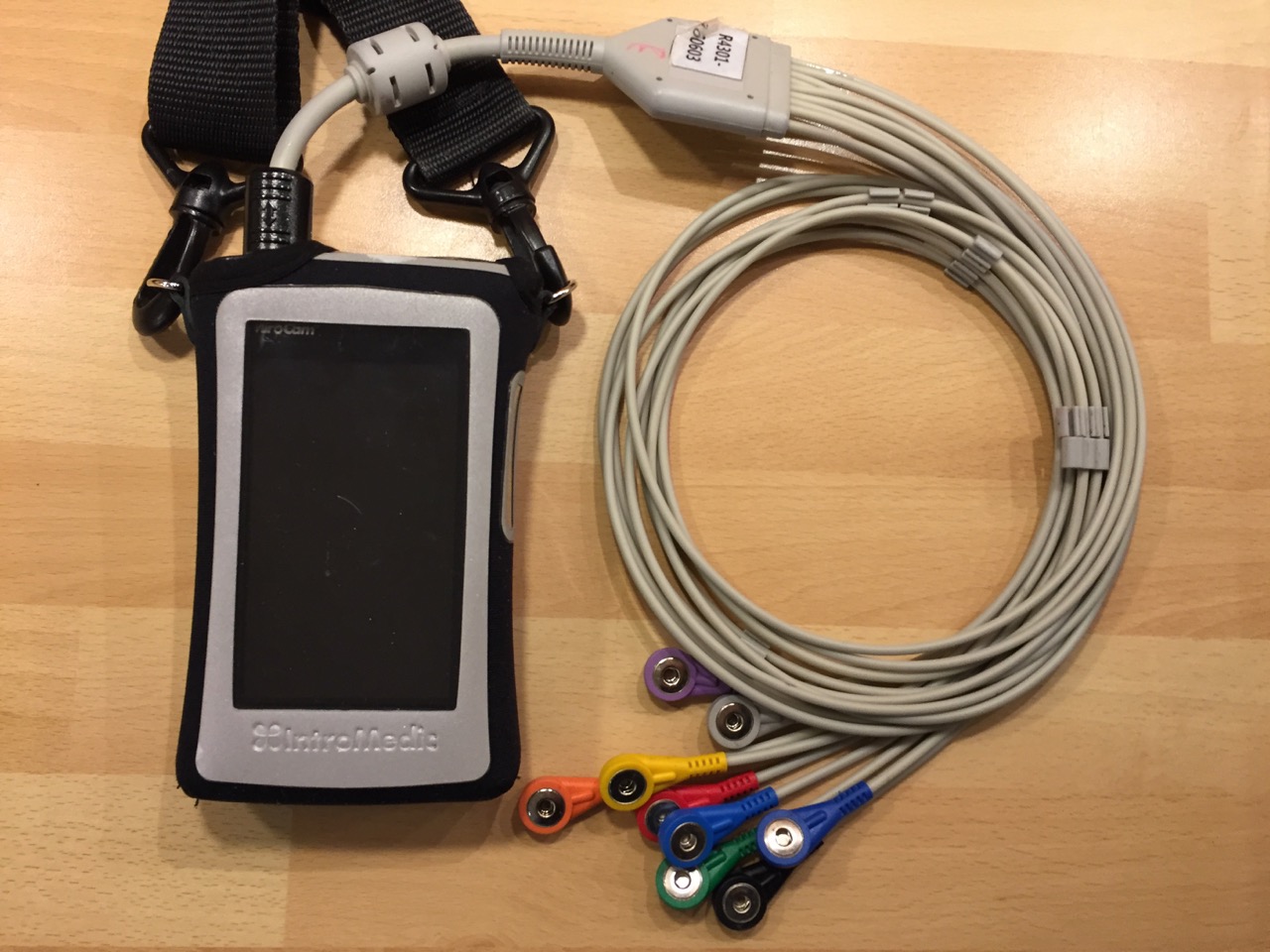
Several manufacturers make capsule systems but they all work on similar principles. There are different cameras for specific tasks, even a double ended one. The more advanced capsules have higher resolutions and frame rates. Some communicate with the recorder via bluetooth, without the need for sensors. Some simply pass through the digestive system and are flushed away; others need to be “recovered” at the end of their journey.
This particular system relied upon an array of sensors to pick up the signal from the capsule and send it to the recorder. The nurse ran through the risks of the procedure. The worst one being the, previously mentioned, capsule becoming stuck and the possible means required to extract it. I signed the consent form.
The first task was to attach the numbered sensors in the correct positions around the abdomen. I could see why wireless communication is the future. (In the interests of good taste I would normally refrain from including a picture of my abdomen, but in this case I thought it worth making an exception)

With the sensors correctly positioned the nurse produced the capsule and asked me to hold it between my finger and thumb then pass it in front of the recorder unit. A bleep showed that they were now “paired”. As she had already input my information into the unit, the display showed my name, hospital number etc.

How easy was it to swallow the capsule? “Very easy. One gulp of water and it was on its way”. The nurse switched on the live monitoring function and we watched it pass down my throat and enter the stomach. To save battery power she then switched the screen off and I didn’t have the courage to try it myself in case I ruined the whole procedure. And what if I had seen something that, to my eyes, looked serious? A surefire way of inducing stress.
The unit had a 12 hour battery life and would switch itself off just before midnight. At that point the sensors could be removed. The equipment would then need to be returned to St.Thomas’. We agreed that I would take it back on the Wednesday. A fortnight later the results should be available.
When would I be able to eat and drink again? Coffee – two hours after swallowing the camera and then a light meal after a further two hours.
If it had been decent weather I would have set off on another walk around London, as light exercise helps the transit of the capsule, but I decided I would rather get home in the warm. I took a short detour to College Green, opposite the Houses of Parliament, to see if there was the usual media village that appears in times of political upheaval. I was surprised to find it empty.
True to the nurse’s word the unit switched itself off precisely 12 hours from the start of the procedure and I was able to peel off the sensors with remarkably little hair loss or pain. Wearing the receiver unit took me back to having a stoma as it was hanging in the same position as a bag and the adhesive on the sensors gave a similar sensation to that of a pouch backplate.
The analysis of the video was due to take 2 weeks nothing was forthcoming. I contacted my gastro consultant who said he would chase it up but after a further 4 weeks there was still no sign of it. Something to quiz the Endoscopy Dept. about at my forthcoming visit.
Tuesday 18th December 2018 – St.Thomas’ – Endoscopy Department
Time for my annual upper GI endoscopy. I had already undergone this procedure at St.Thomas’ ten times before. As with last year’s scoping it was being carried out by the head of the Gastro Department. I asked him whether he could find the results from the capsule endoscopy on the system. He went off to check their status. By the time he returned I had been prepared for the scope – xylocaine spray to back of throat; mouthguard in position; Fentanyl injected. I was unable to speak. Luckily they had held off with the Midazolam so I was, at least, still conscious!
He told me that the video was being checked now but he had seen the first half of it and there appeared to be Crohn’s inflammation in my small intestine. Not what I wanted to hear and with that the Midazolam was injected….zzzzz
I would have to await the full analysis before discussing the way forward. I emailed my gastro consultant to tell him the news and he replied that he would keep an eye out for the report.
Friday 8th March 2019 – St.Thomas’ Hospital – Upper GI Clinic
Another visit to see the surgeon. A surprisingly lovely day and a chance to take as stroll along the South Bank of the Thames.
At my last visit we had discussed removing my gallbladder but agreed to put it on hold until absolutely necessary. I had asked for this new appointment to discuss the mid-right quadrant pain I had been experiencing. I wondered if it was connected to my gallbladder; scar tissue; Crohn’s inflammation; or something else. He had a good prod around and was able to pinpoint the exact centre of the ache. It was very apparent as it hurt when he applied pressure to it.
“You have a small hernia. Usually we would offer you a short operation to repair it but given your history I doubt whether we should consider it” by which he meant the danger of bleeding from the varices in my abdomen outweighed relieving the ache.
Being able to understand and visualise the pain would, at least, make it so much easier to deal with. Should I change my lifestyle? “No, carry on as before but if it gets worse then we will have to revisit the situation. You’ve got our contact number“. Should I consider wearing a support belt as I had done when I had a stoma? “If you feel that it will help“.
I mentioned that I had not yet seen the capsule endoscopy report from last November. He called it up on screen. No wonder they were having difficulties printing it. It comprised page after page of stills from the 12 hour video. There was, however, a summary page and there it was, in black and white, “active inflammation in the proximal (top end of) small bowel and a little further down as well. The far end (anastomosis site) was clear”.
On the train home I mulled over the fact that if it hadn’t been for the elevated calprotectin levels, which prompted the capsule endoscopy, I would have remained blissfully unaware that there was anything wrong. Despite this I was still asymptomatic, had felt nothing and still didn’t. My digestive system was, for most of the time, working as it should. Looking at it now, objectively, the high calprotectin results could only have been either a false positive, albeit a very high and increasing one, or active inflammation in the only section of my digestive tract that hadn’t been seen through a lens.
I didn’t know how close the inflammation had come to causing serious damage or even needing emergency surgery. With the problem identified it was time to find the most suitable treatment and agree how we would measure if it was working.